
Brain Mechanism in MCI Patients during Movie-watching Sub-projects 1
Published in University of Electronic Science and Technology of China, Key laboratory for Neuroinformation of Ministry of Education, 2023
Movie-watching Elicits Memory Recall EEG synchrony connectivity Pattern in People with MCI
Introduction
Aging or aging-related diseases are considered to cause changes in neural synchronization. Natural stimuli such as movie-watching may provide the environment in which recall is evoked. How is neural synchrony affected by MCI disease in this state?
Here we measured the Electroencephalogram (EEG) of 39 normal control (NC) and 33 mild cognitive impairment (MCI) individuals during the viewing of two movies (one of which elicits recall and the other a control) to quantify neural synchrony connectivity and to observe the relationship between changes in synchrony and neuropsychological measures.
In order to select the appropriate movie stimuli for this study, two movies, low-recall and high-recall, were conducted. This was defined based on the subjective screening by the experimental designer, the pre-experiment, and the scale follow-up in the experiment (which mainly depended on the results of the pre-experiment.) The low-recall movie was chosen as a nature documentary; the high-recall movie was a narrative clip whose segment was about going down to the countryside to do farm work. This was due to the fact that the subjects were older Chinese people aged 45-80 years old, and the expectation was that it would elicit memories from the subjects. 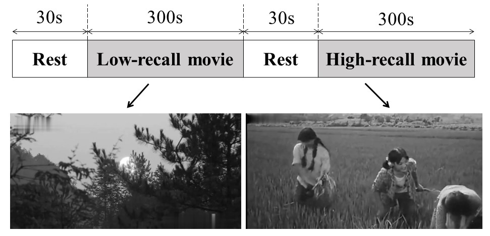
Results
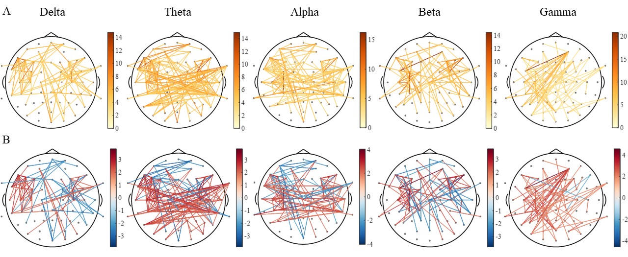
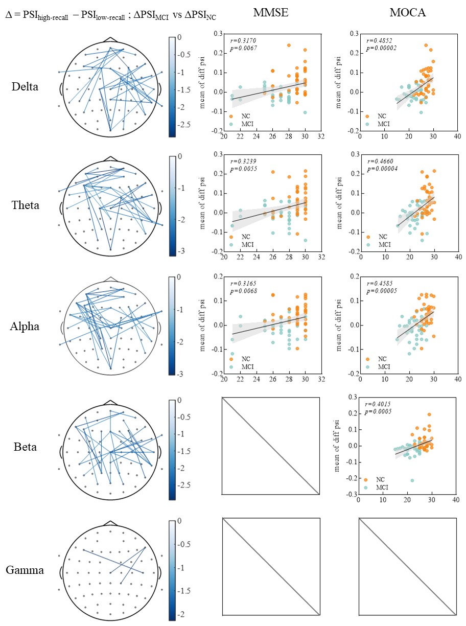
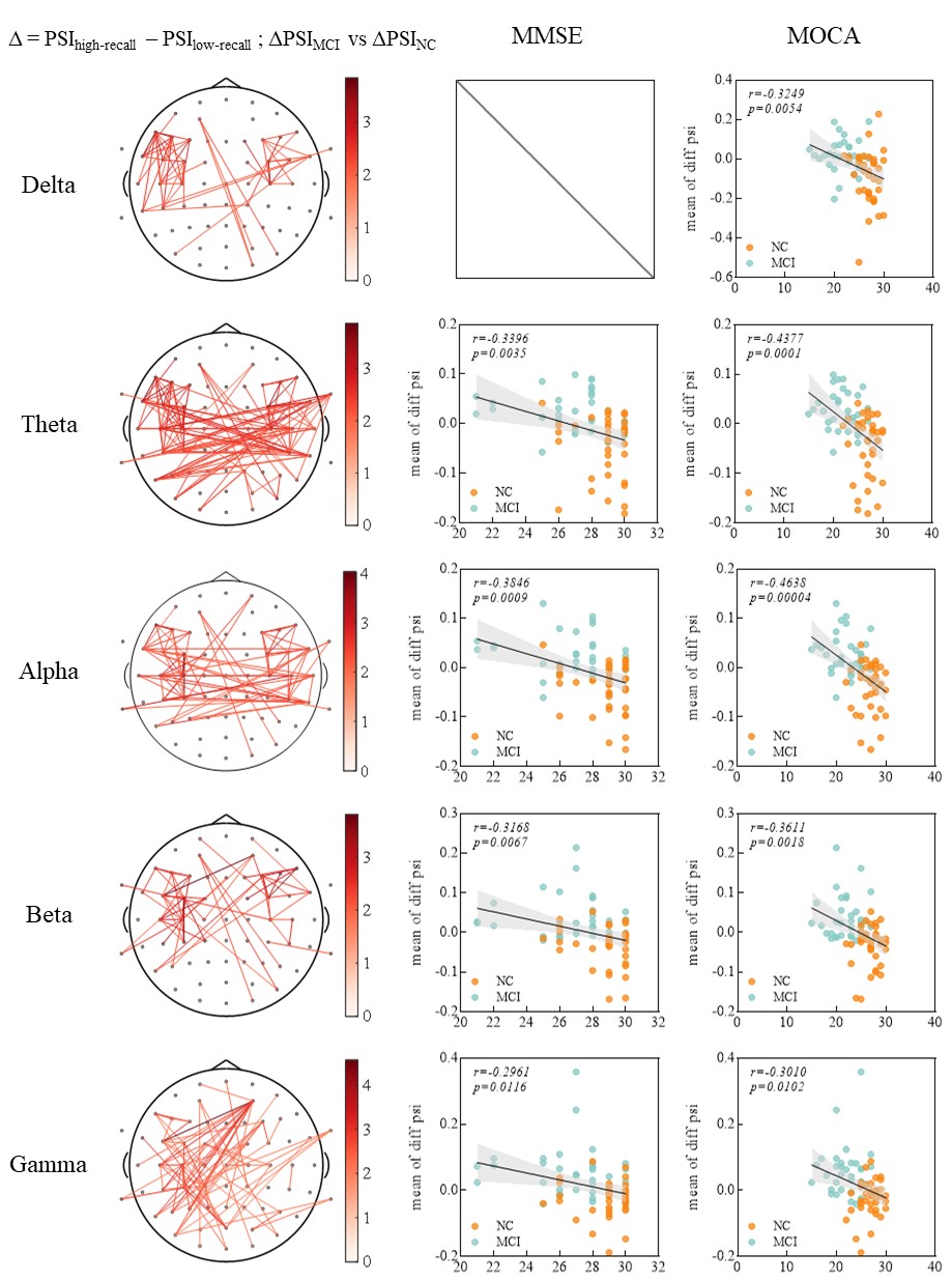
The results showed that the two groups showed different patterns of connectivity distribution in the two states especially in the low frequency bands, with increased synchronization in the frontal-parietal network of the NC and decreased synchronization between the bilateral temporal lobes as the level of recall elicited by the movie increased. Then MCI did not show the same changes, with little change in the frontal-parietal network and abnormal increase in the inter- and inner- temporal lobe network.
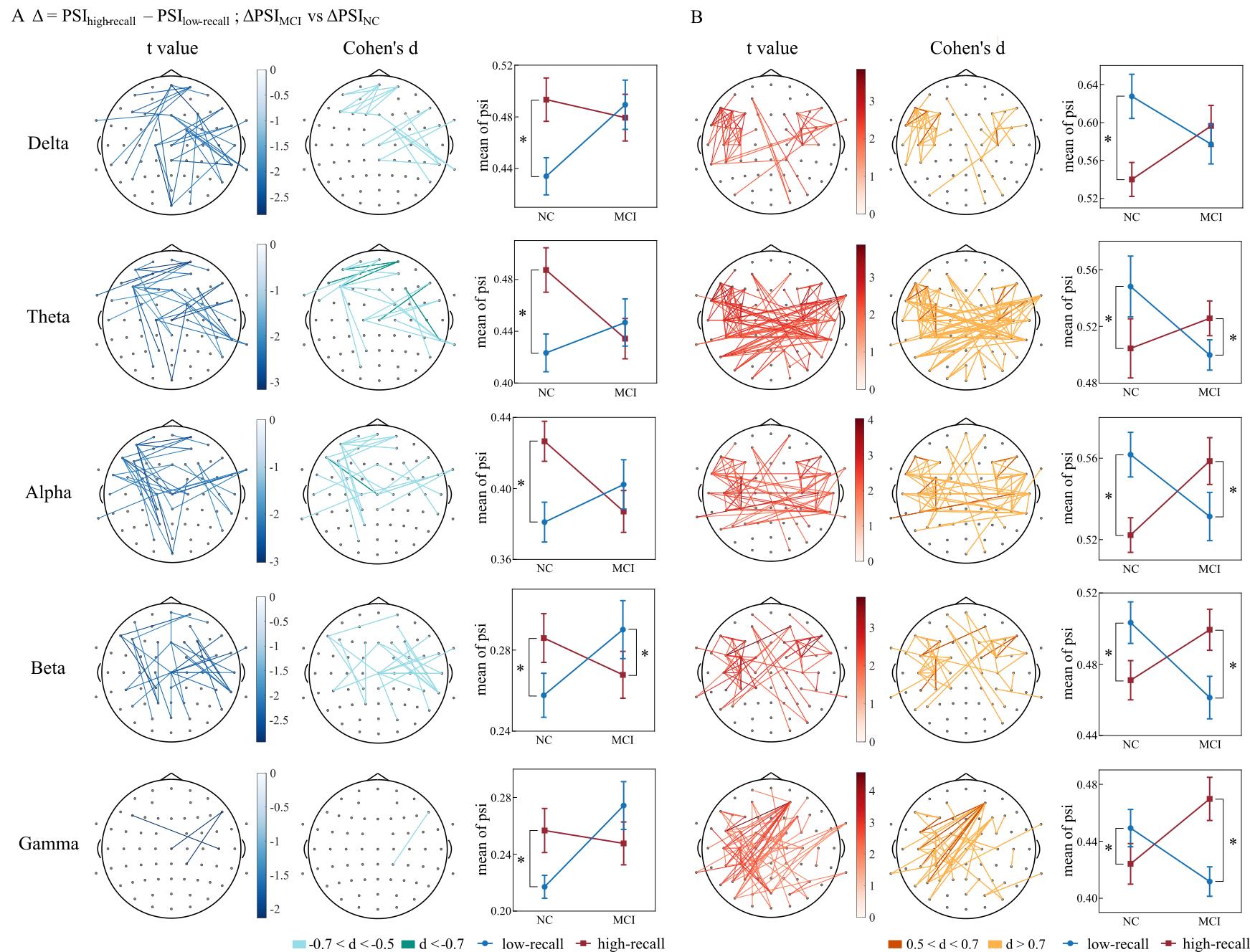
Furthermore, the changes in synchronous connectivity are related to cognitive function which has been suggested by the correlation with neuropsychological assessment scores.
Discussion
Enhancement of FPN during recall in NC and FPN nonresponsiveness to recall in MCI patients, indicated that FPN may play a key role in the recall process. Most studies have demonstrated that prefrontal and parietal regions were activated by many cognitive functions, which play a crucial role in brain information processing as the brain performs higher-level cognitive activities. It showed impaired connectivity between the frontal and parietal brain regions in patients with symptoms of cognitive impairment, especially in MCI patients, the decline in functional connectivity of the frontal-parietal network was detected. Our results may also serve as confirmation of this hypothesis.
The tendency for inter-hemisphere and inner-region connectivity to enhance within the temporal regions of MCI. The brain activity of MCI patients might be activated by movie-watching to produce network changes, for the possession of retrieval memory generated the impairment of the network coordination, and recruited additional neural resources of inter-temporal and inner-temporal networks, which is a disordered network.
Moreover, our finding of between the above difference in connectivity and correlations with neuropsychological measures may reflect the fact that changes in brain network connectivity during the progression of MCI to AD may be a developmentally progressive process.
Limitation
- The recollections in the study we assumed would produce autobiographical recollections, but in reality, movie-watching involves a variety of cognitive activities such as attention, memory retrieval, emotion, and so on. Although it is mentioned that movies provide rich stimuli, the part of the movie stimuli that involves memory retrieval of the stimuli is not isolated (there is no associated psycho-cognitive task), so it is not possible to identify whether the activity of the network is due to memory retrieval processes, but only to indicate that higher cognitive processes are involved. The ability to provide psycho-cognitive tasks in addition to neuropsychological measures would have provided a more accurate experimental group.
- The EEG does not have enough resolution to provide deeper information and needs to be supported by more modal data.
Methods
- Raw EEG data was preprocessed. The details of preprocessing contained: 1) A Hamming window FIR filter was used for 1-40 Hz bandpass filtering to filter out ultra-low frequencies, unwanted high frequencies, and industrial frequency noise; 2) Remove artifact by Independent Component Analysis (ICA) based Multiple Artifact Rejection Algorithm (MARA); 3) Bad channels were identified and interpolated by using reference electrode standardization interpolation technique (RESIT), and re-referencing to REST; 4) Computational platform was used for inspection to identify and remove segments of EEG contaminating excessive noise which with unusually high or low amplitude. Clean EEG data were obtained finally.
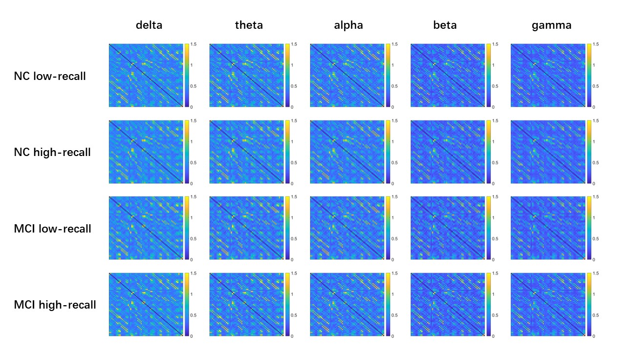
- Phase Synchronization Index (PSI) is calculated to characterize synchronized brain activity.
- Changes in the connectivity of electrodes were characterized across subjects using a two-way mixed (2 groups×2 conditions) analysis of variance (ANOVA) with condition (low-recall vs high-recall movie-watching) as the within-subject factor and group (NC vs MCI) as the between-subject factor in the analysis. A post-hoc t-test were used to investigate potential changes in the interaction factor (p<0.05).
Conclusion
Our study revealed that MCI leads to altered neural synchrony connectivity in the recall state. There are regularities in these network patterns that may be relevant to cognitive and recall functions.